
Anatomy Of The Foot Bottom Anatomy Of The Bottom Of The Foot Human
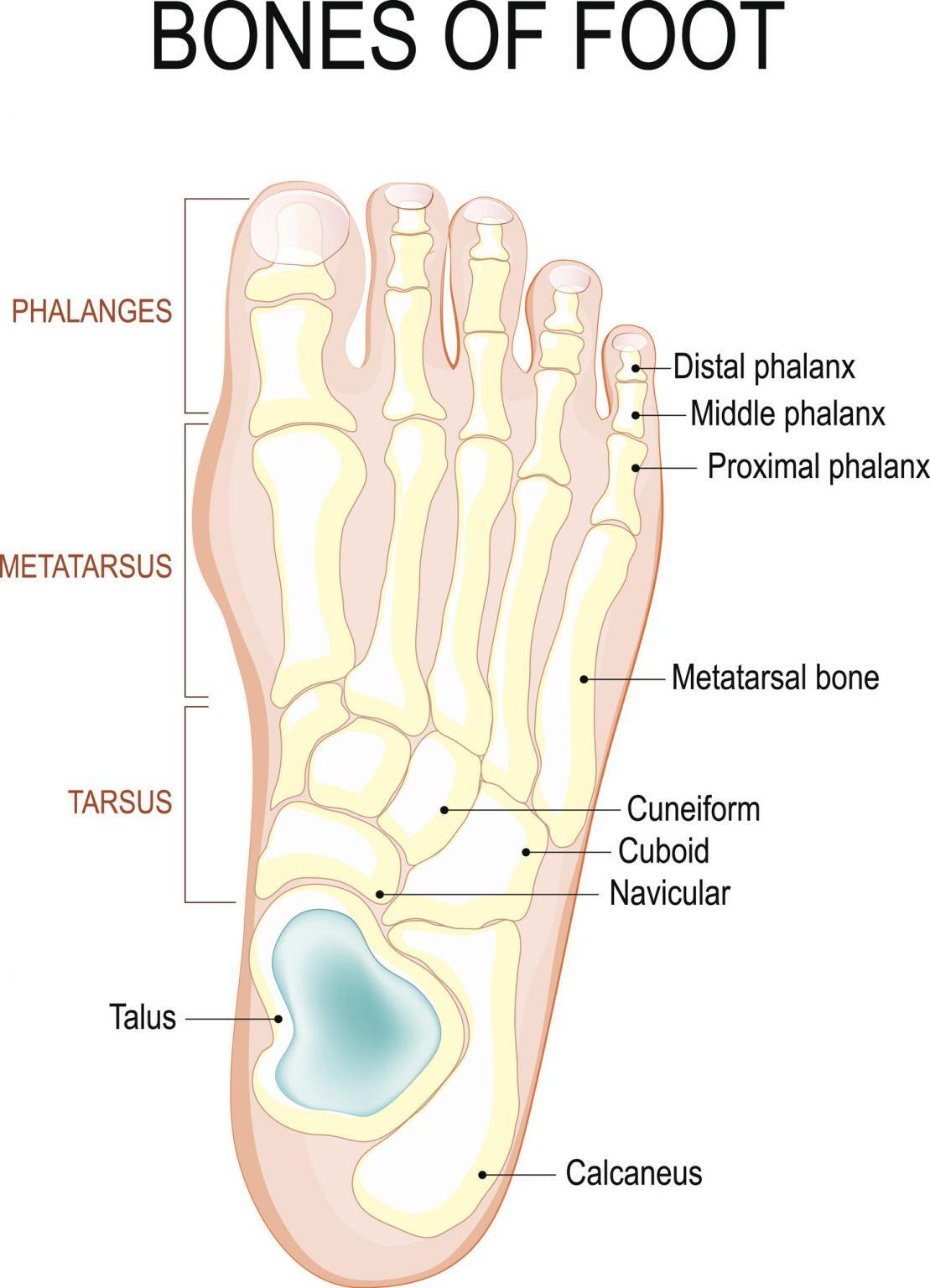
The ankle joint is a hinge joint that allows for dorsiflexion (bending up) and plantarflexion (bending down) of the foot. Sesamoid Bones There are two of these small, ovoid-shaped bones located beneath the first metatarsal on the plantar (surface) of the foot. It's embedded in a tendon at the head (the closest part to the big toe) of the bone.

Diagram Of Your Foot
The bottom of your foot is connected with your pelvic area. Even the basics can lead you to a very simple self-treatment. The right foot is associated with the right part of the body and left foot is associated with the left side of the body.
Plantar
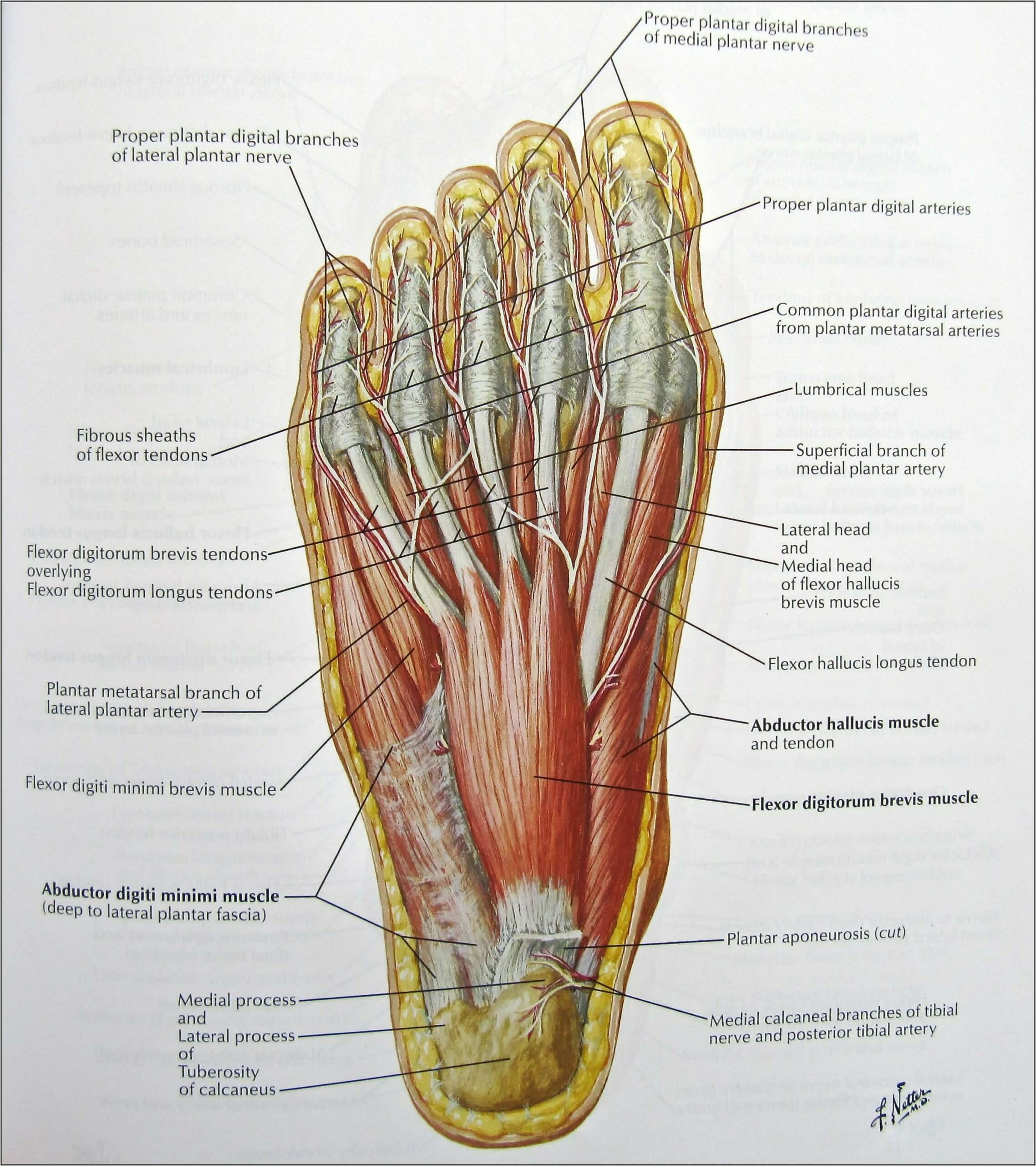
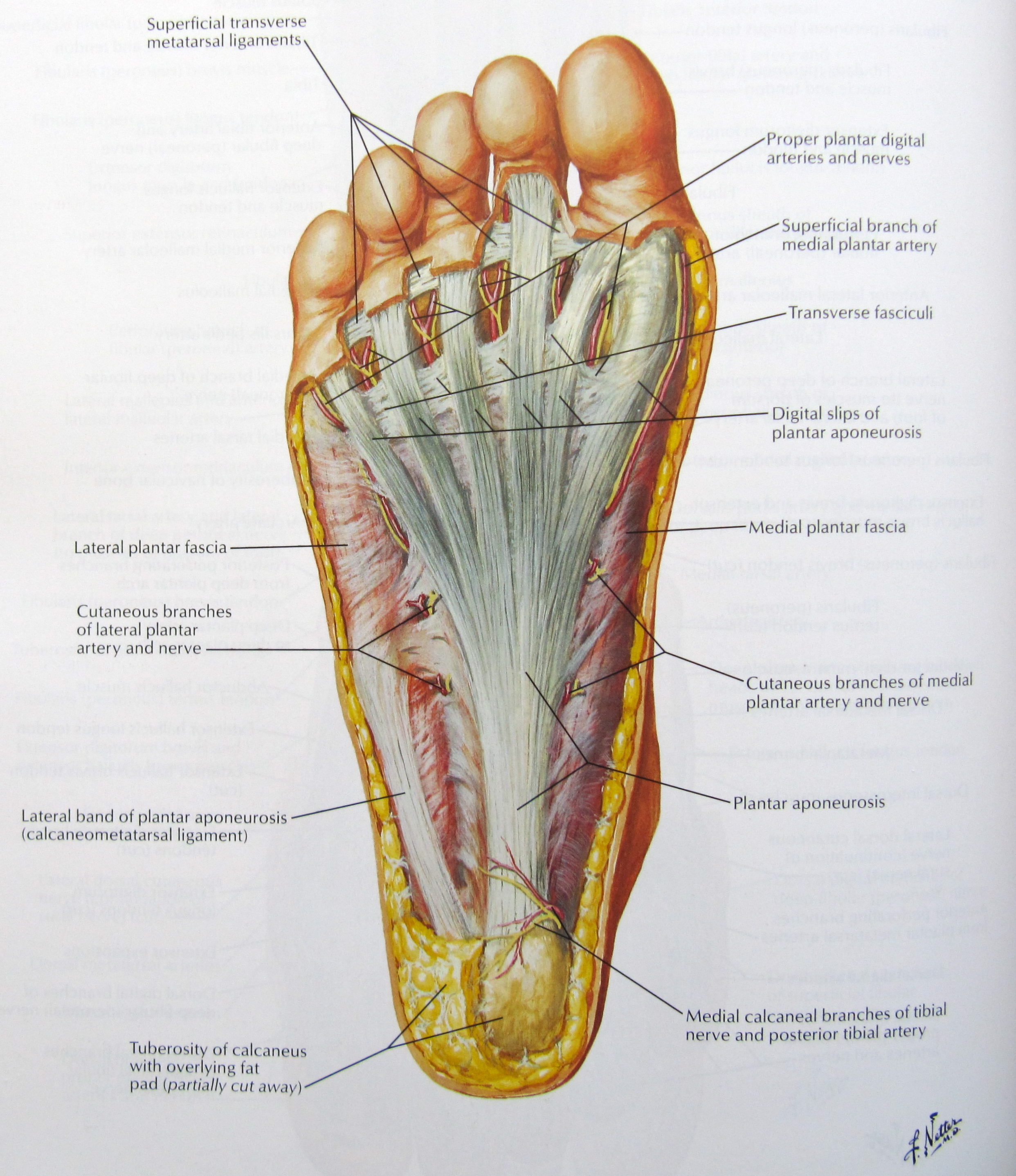
LABELED DIAGRAMS. Figure 1. Sections and Bones of the Foot A. Lateral (Left) B. Anterior (Right) Figure 2. Compartments of the Foot A. Cut Section through Mid-Foot. Figure 3. First Layer of the Foot A. Plantar View of Right Foot. Figure 4. Second Layer of the Foot A. Plantar View of Right Foot.

Anatomy of the Foot Comprehensive Orthopaedics
Tragedy may have been averted Friday night when a panel of a Boeing plane blew out as an Alaska Airlines flight traveled at 16,000 feet, an NTSB official said Saturday night.. Seats adjacent to.
Anatomy Regions Of The Right Foot Wood Print by Asklepios Medical Atlas
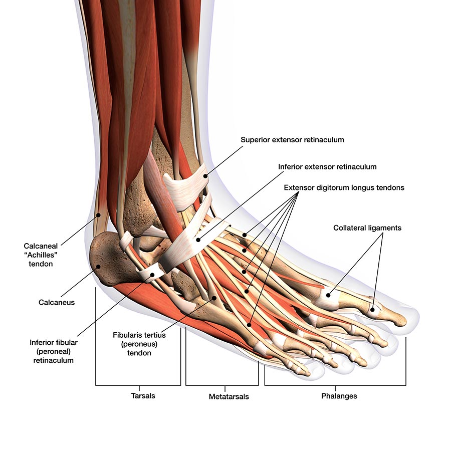
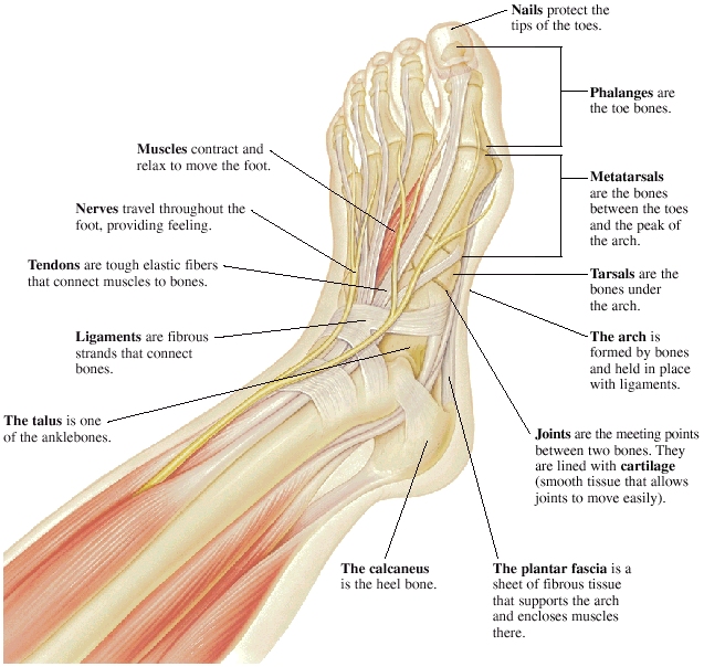
Nails protect the tips of the toes. Phalanges are the toe bones. Metatarsals are the bones between the toes and the ankle bones. Tarsals are bones of the rear foot (hindfoot) or middle foot (midfoot). The talus is one of the ankle bones. The calcaneus is the heel bone. The arch is formed by bones and held in place with ligaments.

The bones in the foot inferior view (Picture illustrated from Thieme
The midfoot is a pyramid-like collection of bones that form the arches of the feet. These include the three cuneiform bones, the cuboid bone, and the navicular bone. The hind foot forms the heel and ankle. The talus bone supports the leg bones (tibia and fibula), forming the ankle. The calcaneus (heel bone) is the largest bone in the foot.
Foot and ankle anatomy, conditions and treatments
The outsole of the foot is the part on the bottom of the shoe that touches the ground. A softer sole provides greater ability to absorb shock. The bottom, back part of the shoe is called the heel. The heel gives the shoe elevation. A higher heel places more pressure on the balls of the feet and toes.

Muscle Anatomy Of The Plantar Foot Everything You Need To Know Dr
The hindfoot This is comprised of the talus bone and the calcaneum. The talus connects with the tibia and fibula to form the ankle joint, and the calcaneum is the bone that forms the heel bone (ball of the foot). The calcaneum is the largest bone in the foot. The muscles, tendons and ligaments
Foot, Parts of Anatomy and Physiology
Introduction A solid understanding of anatomy is essential to effectively diagnose and treat patients with foot and ankle problems. Anatomy is a road map. Most structures in the foot are fairly superficial and can be easily palpated. Anatomical structures (tendons, bones, joints, etc) tend to hurt exactly where they are injured or inflamed.
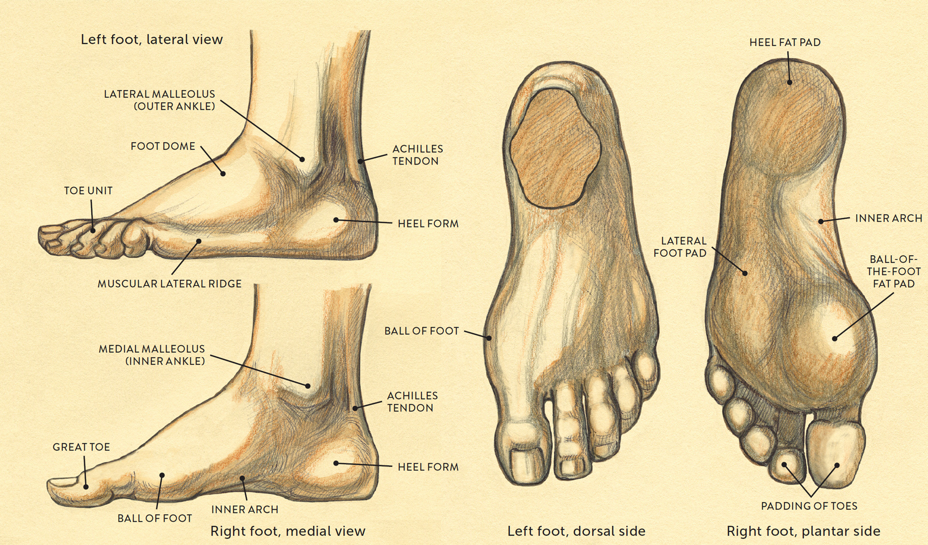
SURFACE FORM LANDMARKS OF THE FOOT
Summary The foot is an intricate part of the body, consisting of 26 bones, 33 joints, 107 ligaments, and 19 muscles. Scientists group the bones of the foot into the phalanges, tarsal bones,.
Medical Diagram Of Bottom Of Foot Diagrams Resume Template
The bottom part of the foot is the sole. The padded area on the bottom of the foot is known as the plantar aspect. The top part of your foot above the arch is the instep. In medical terms, the top of the foot is the dorsum or dorsal region. Anatomy of the Lower Leg Muscles. Bones .
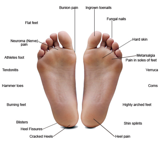
Common Foot Problems — Hawaii Podiatry
It is made up of over 100 moving parts - bones, muscles, tendons, and ligaments designed to allow the foot to balance the body's weight on just two legs and support such diverse actions as running, jumping, climbing, and walking. Because they are so complicated, human feet can be especially prone to injury.
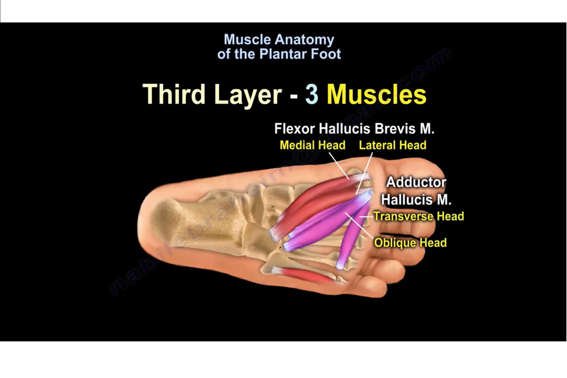
Muscle Anatomy Of The Plantar Foot —
Rest Cold packs Anti-inflammatory medication, such as ibuprofen Proper stretching before activity Proper footwear or shoe inserts Corticosteroid injections Surgery (for more severe, prolonged conditions) What is a corn? Corns are yellowish, callus growths that develop on top of the toes. Corns develop because of abuse or stress.
Notes on Anatomy and Physiology Using Imagery to Relax the Weight
Generally, the three groups are the: tarsals metatarsals phalanges Tarsals The tarsals are a group of seven bones close to the ankle. The proximal tarsal bones are the talus and the calcaneus,.

Cutaneous afferent innervation of the human foot sole what can we
Ball-of-foot pain. Pain in the ball of the foot, or at the front of the foot near the toes, has many potential causes. Muscle strains and sprains, minor overuse injuries, and tense muscles can all.
What is Metatarsalgia? JOI Jacksonville Orthopaedic Institute
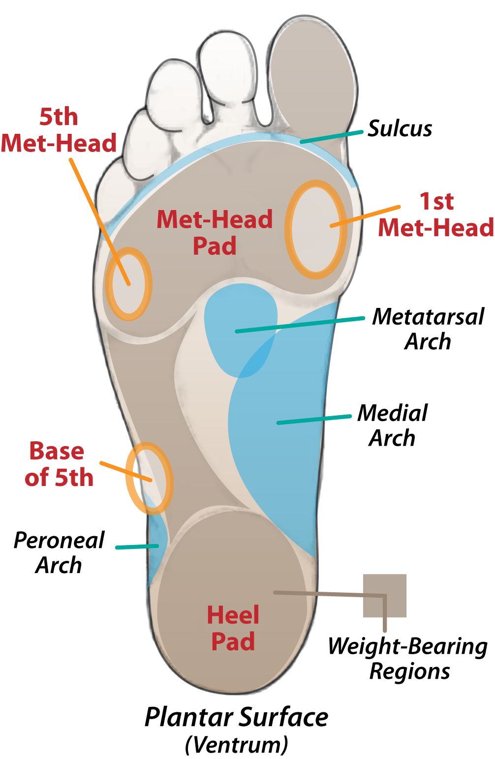
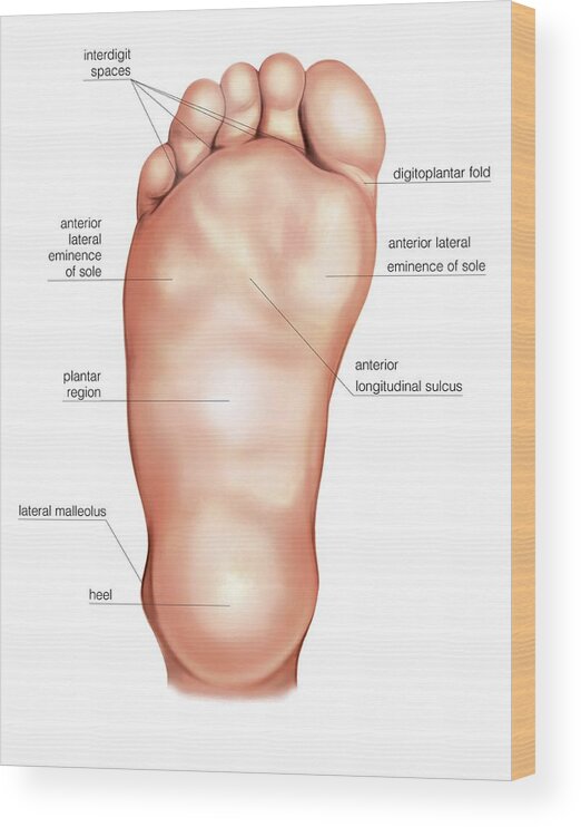
The sole is the bottom of the foot . In humans the sole of the foot is anatomically referred to as the plantar aspect . Structure Deep anatomy of the sole The glabrous skin on the sole of the foot lacks the hair and pigmentation found elsewhere on the body, and it has a high concentration of sweat pores.